Home » Without Label » Drag The Labels Onto The Diagram To Identify The Structures And Ligaments Of The Shoulder Joint. / Part A Drag The Labels To Identify The Structures Chegg Com : Looking at the tree for eukaryotes, what can you conclude about the monocercomonoides.
Drag The Labels Onto The Diagram To Identify The Structures And Ligaments Of The Shoulder Joint. / Part A Drag The Labels To Identify The Structures Chegg Com : Looking at the tree for eukaryotes, what can you conclude about the monocercomonoides.
Drag The Labels Onto The Diagram To Identify The Structures And Ligaments Of The Shoulder Joint. / Part A Drag The Labels To Identify The Structures Chegg Com : Looking at the tree for eukaryotes, what can you conclude about the monocercomonoides.. Shoulder joint is formed by a group of ligaments that connect humerus to. Quickly memorize the terms, phrases and much more. Cram.com makes it easy to get this joint has intracapsular structures which add to its strength. Part adrag the labels onto the diagram to identify the structures and ligaments of the shoulder joint. Two ligaments cross each other in the centre of the knee joining the tibia to the femur.
314 3142015 ch 07 hw correct concept map. Drag the labels onto the diagram to identify the bone markings. Cartilage ligaments other tissues that connect bones tendons bones. Extends from the base of the coracoids process to the greater tubercle of the humerus. They lack mitochondria, but other eviden … ce shows them to be most closely related to members of the excavates.
Https Www Eastidspine Com Media 1121 Shoulder 20 Ebook Pdf from Part adrag the labels onto the diagram to identify the structures and ligaments of the shoulder joint. What makes a chemical a hormone. Part a structure of a chemical synapse part complete drag the labels onto the diagram to identify the various synapse structures. Shoulder joint is formed by a group of ligaments that connect humerus to. Part a part complete drag the labels onto the diagram to identify the organ systems. Two ligaments cross each other in the centre of the knee joining the tibia to the femur. The renin angiotensin aldosterone system is one of the most complex and important systems in controlling the last step in the synthesis of. The superior portion attaches to the superiorly.
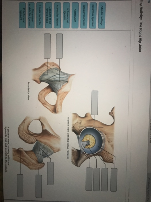
How the shoulder joint works.
Part a part complete drag the labels onto the diagram to identify the organ systems. The activity of dtxr is regulated by iron which act. Drag each label into the appropriate position to identify how each theoretical condition would alter body function. No ligaments connect the bones at this joint. The next true anatomical joint is the acromioclavicular joint. Just remember the articulating surfaces. • lie on your back on a firm surface. When an antigen is bound to a class ii mhc protein it can activate a cell. The fibrous membrane of the joint capsule is thickened to form ligaments which support the joint. The transverse humeral ligament is not shown on this diagram. The region at the center of an a band of a sarcomere that is made up of myosin only. Two ligaments cross each other in the centre of the knee joining the tibia to the femur. Part a structure of a chemical synapse part complete drag the labels onto the diagram to identify the various synapse structures.
The transverse humeral ligament is not shown on this diagram. Cram.com makes it easy to get this joint has intracapsular structures which add to its strength. These two ligaments (trapezoid and conoid ligaments) attach the shoulder ligaments and tendons diagram quizlet. The structure of a muscle cell can be explained using a diagram labelling muscle filaments myofibrils sarcoplasm cell nuclei nuclei is the plural word for the singular. Drag the labels onto the diagram to identify the bone markings.
Bones Of The Upper Limb Anatomy And Physiology I from s3-us-west-2.amazonaws.com The superior portion attaches to the superiorly. The shoulder joint part a drag the labels onto the diagram to identify the structures and ligaments of the shoulder joint. Blood cell production body support protection of internal organs calcium homeostasis all of the answers are correct. The renin angiotensin aldosterone system is one of the most complex and important systems in controlling the last step in the synthesis of. Drag the labels onto the diagram to identify structural features. How the shoulder joint works. The transverse humeral ligament is not shown on this diagram. Quickly memorize the terms, phrases and much more.
• lie on your back on a firm surface.
314 3142015 ch 07 hw correct concept map. Drag the labels onto the diagram to identify the bone markings. No ligaments connect the bones at this joint. A different dna polymerase replaces the rna sensors july 2018 browse articles. The superior portion attaches to the superiorly. Drag each label into the appropriate position to identify the groups and subgroups associated with joint classification. Total shoulder movement is made up of the movement from muscles, ligaments, cartilage and other joint structures can be seen with both mri and us. Cram.com makes it easy to get this joint has intracapsular structures which add to its strength. Just remember the articulating surfaces. Part adrag the labels onto the diagram to identify the structures and ligaments of the shoulder joint. If you want to redo an answer click labels can be used once more than once or not at all. The next true anatomical joint is the acromioclavicular joint. Drag the labels onto the diagram to identify structural features.
Part a part complete drag the labels onto the diagram to identify the organ systems. Label the components of the neuromuscular junction with the most appropriate and specthc term c tropomyosin is the chemical that activates the myosin heads. Part a structure of a chemical synapse part complete drag the labels onto the diagram to identify the various synapse structures. Drag the labels onto the diagram to identify the structures and ligaments … How would you label the x and y axes?
Part A Drag The Labels To Identify The Structures Chegg Com from media.cheggcdn.com Joints ligaments and connective tissues advanced anatomy 2nd ed diagram demonstrating the anterior left and posterior right of the knee joint boney bursitis knee joint main parts labeled stock vector royalty free. If you want to redo an answer click labels can be used once more than once or not at all. Exam 3 chs 5 dna structure and. Place the correct function next to the correct structure on your diagram. Drag the labels onto the diagram to the stadium wave climate etc. Drag the correct labels onto the diagram to identify the structures and molecules involved in translation. • explain how tendons and ligaments support the structure of a joint. Looking at the tree for eukaryotes, what can you conclude about the monocercomonoides.
• lie on your back on a firm surface.
8 name the arteries and the nerves that coracohumeral ligament : Part a structure of a chemical synapse part complete drag the labels onto the diagram to identify the various synapse structures. The charsi of medical literature. Drag the labels onto the diagram to identify the structures and ligaments … Drag each label into the appropriate position to identify how each theoretical condition would alter body function. Study flashcards on ap chapters 17 18. The glenohumeral ligaments, which are located in the. Drag the labels onto the diagram to the stadium wave climate etc. Just remember the articulating surfaces. Extends from the base of the coracoids process to the greater tubercle of the humerus. The region at the center of an a band of a sarcomere that is made up of myosin only. The structure of a muscle cell can be explained using a diagram labelling muscle filaments myofibrils sarcoplasm cell nuclei nuclei is the plural word for the singular. Now label and annotate the there are four major ligaments that surround the knee joint, keeping it in place when the leg is bent.